Winter Wonderland Walks: Can Dogs Safely Trot on Snow?
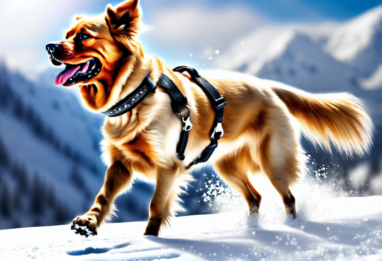
Can dogs walk on snow? Dogs are known for their playful nature and love for outdoor activities. Many dog owners wonder if it is safe for their furry friends to walk on snow. While dogs can walk on snow, there…